Special offer! Comprehensive professional teeth cleaning for only 1699 UAH.
Special offer! Comprehensive professional teeth cleaning for only 1699 UAH.
Special offer! Comprehensive professional teeth cleaning for only 1699 UAH.
Special offer! Comprehensive professional teeth cleaning for only 1699 UAH.
Special offer! Comprehensive professional teeth cleaning for only 1699 UAH.
Special offer! Comprehensive professional teeth cleaning for only 1699 UAH.
Special offer! Comprehensive professional teeth cleaning for only 1699 UAH.
Special offer! Comprehensive professional teeth cleaning for only 1699 UAH.
Special offer! Comprehensive professional teeth cleaning for only 1699 UAH.
Special offer! Comprehensive professional teeth cleaning for only 1699 UAH.
пр.Лобановського, 130, Київ, Україна
Temporarily closed
Opening soon

78A, Irpinska St., Kyiv, Ukraine
Temporarily closed
Opening soon

32A, Heroiv Dnipra St., Kyiv, Ukraine
Temporarily closed
Opening soon

9B, Yevhena Chykalenko St. (Pushkinska), Kyiv, Ukraine
Temporarily closed
Opening soon

General Dentistry
Dental surgery
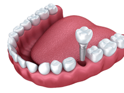
Dental Implants
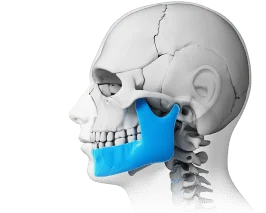
Maxillofacial Surgery
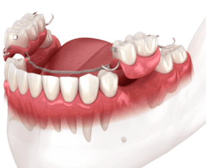
Dental prosthetics
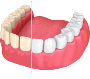
Cosmetic dentistry
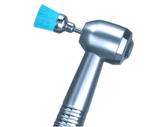
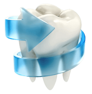
Professional Dental Hygiene

Periodontics
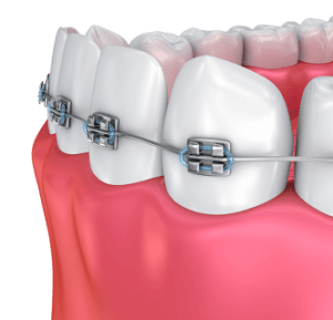
Orthodontic dentistry
Gnathology & TMJ Therapy

Pediatric Dentistry
Dental X-Rays
Popular requests
Teeth whitening
Treatment of caries
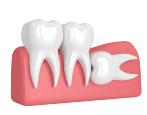
Wisdom tooth extraction
Bracket system
Dental veneers
Tooth restoration
Pediatric dentist
Consultation with a dentist-therapist
Search results by: «».
Unfortunately, no results were found for your search: “Pediatric Endocrinologist”, nothing was found.
You may find these links useful:
Clinics "MED-DEO"
пр.Лобановського, 130, Київ, Україна
Temporarily closed
Opening soon
78A, Irpinska St., Kyiv, Ukraine
Temporarily closed
Opening soon
32A, Heroiv Dnipra St., Kyiv, Ukraine
Temporarily closed
Opening soon
9B, Yevhena Chykalenko St. (Pushkinska), Kyiv, Ukraine
Temporarily closed
Opening soon
Services
DentistryGeneral Dentistry
Dental surgery
Dental Implants
Maxillofacial Surgery
Dental prosthetics
Cosmetic dentistry
Professional Dental Hygiene
Periodontics
Orthodontic dentistry
Gnathology & TMJ Therapy
Pediatric Dentistry
Dental X-Rays

")
Service statistics

More and more clients choose «MED-DEO»

Initial consultation with a doctor
The cost of a consultation: 1000 UAH.
Appointment by phone:
(044) 394 90 94An atheroma (epidermoid cyst) is a benign subcutaneous formation that appears due to blockage of the sebaceous gland duct.
The good news is that atheroma can be easily removed using modern methods without serious consequences for health and skin aesthetics.
Atheroma Removal - Price 2026
Service name
Price, UAH.
Первинна консультація щелепно-лицевого хірурга кандидата медичних наук
1200 ₴
Sign upПовторна консультація щелепно-лицевого хірурга кандидата медичних наук
800 ₴
Sign upПервинна консультація щелепно-лицевого хірурга
800 ₴
Sign upПовторна консультація щелепно-лицевого хірурга
600 ₴
Sign upЛікування під седацією
from 5800 ₴
Sign upКТ верхньої та нижньої щелепи
1200 ₴
Sign up")

Reviews
Causes of Atheroma: Why Sebaceous Glands Become Blocked
The main cause of atheroma is blocked sebaceous glands. When sebum cannot exit to the surface, it accumulates, stretches the duct, and forms a capsule—this is how a subcutaneous formation occurs. Main reasons why sebaceous glands can become blocked:
- Increased sebum production—oily skin, seborrhea, or hormonal changes during adolescence, pregnancy, menopause, or with endocrine disorders.
- Changes in sebum composition—secretion becomes thicker and cannot pass through the duct.
- Acne and skin inflammation—with acne, ducts can scar or become deformed.
- Excessive sweating (hyperhidrosis)—disrupts skin balance and contributes to blockage.
- Mechanical skin damage—cuts, abrasions, boils, or scars can interfere with normal gland function.
- Poor hygiene—accumulation of dirt, cosmetic residue, and dead cells can clog pores.
- Genetic predisposition—structural and functional features of sebaceous glands can be inherited.
- Hormonal factors—elevated androgen levels stimulate sebum production.
- Metabolic disorders—diabetes, obesity, and other metabolic changes increase risk.
- Poor-quality cosmetics—comedogenic products block pores and trigger cyst formation.
So, atheroma occurs due to a combination of internal and external factors, from hormonal changes to skin characteristics and lifestyle. Despite this, it can be easily managed: modern atheroma removal methods are safe and effective, allowing you to avoid complications and maintain skin health.
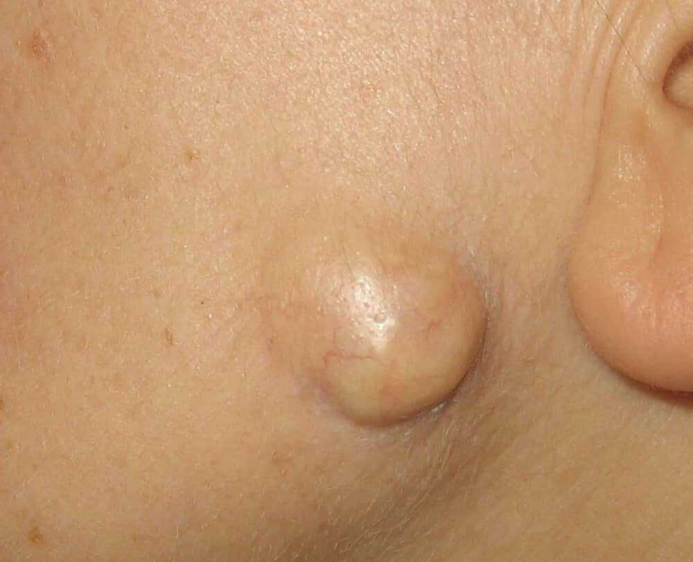
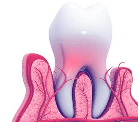
What Atheroma Looks Like: Symptoms and Visual Signs
Atheroma is a subcutaneous formation that’s easy to spot with the naked eye. What does atheroma look like? Visually, it usually has a round or oval shape with clear boundaries and protrudes slightly above the skin. When touched, atheroma is elastic, soft, and mobile under the skin—you can easily move it with your fingers.
Atheroma size varies from 0.5 cm to 5–7 cm, with cysts of 1–3 cm being most common. A typical sign is a small dark spot in the center, corresponding to the blocked sebaceous gland pore.
When pressed, atheroma may release thick whitish-yellow contents with a characteristic unpleasant odor. Self-squeezing is strictly prohibited, as it increases the risk of infection and complications.
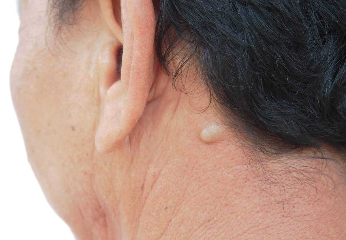
In most cases, atheroma doesn’t hurt and causes no discomfort. The main problem is a cosmetic defect, especially if the cyst is located on the face, neck, or exposed areas of the body. Photos of atheroma help identify typical locations and sizes: cysts most often appear on the face, neck, back, and back of the head.
If atheroma starts to hurt, redden, increase in size, or swelling appears—these are signs of inflammation. In such cases, immediate medical consultation is needed to avoid complications and quickly remove the cyst without harming the skin. Often, infected atheroma looks like the photo below.

Where Atheroma Most Often Appears: Localization

Atheromas form only where sebaceous glands exist, meaning practically anywhere on the body except palms and soles. They most often occur in these locations:
- Scalp (40–50%)—back of the head, temples, parietal area. This is where atheroma on the head or “hard lump on the head” is often noticed. In this area, atheromas can be multiple and small.
- Behind the ear and on the ear (20–25%)—a very common location. Search queries like “atheroma behind the ear” or “ear atheroma” reflect how frequently cysts appear here.
- Face (15–20%)—chin, cheeks, forehead, eyebrow ridges, nasolabial triangle. Facial atheromas attract attention due to cosmetic defects, even if they don’t hurt.
- Back (10–15%)—most often the interscapular area and lower back.
- Neck (5–10%)—back and lateral surfaces of the neck.
- Underarms—less common, but also possible.
- Groin area—quite rare, but also a potential site.
Understanding typical locations helps quickly recognize atheroma and assess whether you should see a doctor for removal.
Is Atheroma Cancer or Not? Debunking the Most Common Myth
Many patients worry about the question “is atheroma cancer?” The answer is simple and unequivocal: atheroma is not cancer. It’s a benign subcutaneous cyst that doesn’t transform into a malignant tumor and doesn’t threaten life. Why confusion arises:
- Atheroma can externally resemble other subcutaneous formations—lipoma, fibroma, or other tumors.
- When inflamed, the cyst enlarges, which sometimes resembles rapid tumor growth.
- Some malignant skin formations (basal cell carcinoma, melanoma) sometimes have a similar appearance.
How to distinguish atheroma from skin cancer:
- Atheroma: soft, mobile under the skin, with clear boundaries, central dark spot, grows slowly over years, painless if not inflamed.
- Skin cancer: firm, fixed to underlying tissues, with unclear boundaries, may ulcerate, grows rapidly, potentially metastasizes.
Even if signs resemble atheroma, with any suspicious skin growth, you should consult a dermatologist or oncologist for accurate diagnosis.
Can Atheroma Go Away on Its Own Without Treatment?
A common patient question—can atheroma go away on its own or disappear without treatment. The short and honest answer: no, atheroma doesn’t go away on its own. Even if it doesn’t change or bother you for a long time, it doesn’t mean the problem has disappeared. Why atheroma can’t resolve itself:
- The atheroma capsule is a formed connective tissue shell that doesn’t disappear without intervention.
- The cyst contents (keratin and sebum) have no exit due to the blocked duct.
- The body cannot independently eliminate such formations.
What happens to atheroma without treatment:
- It can remain unchanged for years, maintaining only a cosmetic defect.
- It may slowly increase in size due to content accumulation.
- It may become inflamed or infected, requiring urgent treatment.
- It may spontaneously rupture, but the capsule remains—and atheroma reappears after a few months.
Sometimes you can find advice about treating atheroma with ointments or anti-inflammatory agents. It’s important to understand—the effectiveness of conservative treatment is not proven. The only way to completely get rid of atheroma is surgical removal along with the capsule. If you remove only the contents, the recurrence risk is 80–90%.

What to Do If Atheroma Ruptures or Becomes Infected: Emergency Care
If atheroma suddenly enlarges, starts hurting, reddens, or shows signs of infection, this means the process has entered an inflammatory stage. In this situation, you can’t delay, because inflamed atheroma is no longer just a subcutaneous formation, but an acute infectious process. Most often, inflammation is accompanied by throbbing or distending pain, tissue tension, local skin temperature increase, swelling, and sometimes general weakness or fever.
Atheroma infection usually occurs due to bacterial invasion, most often staphylococcus or streptococcus. This can happen due to skin microtrauma, decreased immunity, or, most commonly, attempts to squeeze or pierce the formation yourself. Self-treatment in such cases only worsens the situation and increases the risk of infection spreading deep into tissues.
Without timely medical care, infected atheroma can lead to serious complications—abscess formation, phlegmon, tissue necrosis, and in severe and neglected cases, even sepsis. That’s why when pain, redness, or rapid growth of the formation appears, you need to see a surgeon as soon as possible.
Before visiting a doctor, it’s important not to make mistakes. Don’t apply heat, as it accelerates bacterial multiplication and intensifies inflammation. It’s strictly forbidden to try to squeeze or pierce atheroma yourself. You may apply cold through fabric for 10–15 minutes to reduce pain and swelling, and treat the skin with antiseptic if it’s not damaged.
Sometimes atheroma ruptures spontaneously, and purulent contents come out. It’s important to understand this doesn’t mean recovery. In this situation, you need to treat the wound with antiseptic, apply a sterile dressing, and see a surgeon as soon as possible. Spontaneous rupture doesn’t solve the problem, because the atheroma capsule remains under the skin, and eventually the formation reappears.
You should also know that during acute inflammation, it’s impossible to completely remove atheroma. First, the doctor relieves inflammation and infection, and only after that is planned surgical removal of the capsule performed. This approach allows avoiding complications and significantly reduces the risk of atheroma recurrence.
Atheroma Diagnosis: How Doctors Determine the Diagnosis
Atheroma diagnosis is usually not difficult and in most cases doesn’t require lengthy or expensive examinations. An experienced dermatologist or surgeon can suspect atheroma during the initial examination, paying attention to the characteristic appearance of the formation, its typical location, and the presence of a central dark spot—the blocked sebaceous gland duct. This sign often helps distinguish atheroma from other subcutaneous growths.
During examination, the doctor necessarily performs palpation. They assess how mobile the formation is under the skin, what consistency it has, and whether it causes pain. In most cases, atheroma is soft or elastic, moves well, and doesn’t hurt if there’s no inflammation. If pain, redness, or swelling appears, this may indicate atheroma infection, which affects further treatment tactics.
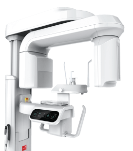
To clarify the atheroma diagnosis and exclude other diseases, the doctor may order soft tissue ultrasound. Ultrasound examination allows visualization of a cystic formation with clear boundaries, presence of a capsule, depth of location, and connection with surrounding tissues. Ultrasound helps distinguish atheroma from lipoma, fibroma, enlarged lymph node, or other subcutaneous formations that may have a similar external appearance.
In some cases, the doctor may use dermatoscopy—examination of skin under magnification. This study is necessary to exclude malignant skin tumors, particularly melanoma, as some growths in early stages may look benign.
After atheroma removal, histological examination is mandatory. The removed formation is sent for pathomorphological analysis, which allows final confirmation of the atheroma diagnosis and exclusion of malignancy. Histology results are usually ready within 7–14 days. Biopsy as a separate diagnostic method is used rarely and only when there are doubts about the nature of the formation. Most often, atheroma is differentiated from the following conditions:
- Lipoma (fatty tumor), which is usually softer and has no central spot
- Fibroma, which is firmer and less mobile
- Hygroma in joint areas
- Lymphadenitis or dermoid cyst
Accurate atheroma diagnosis allows proper treatment method selection, timely removal of the formation, and minimizes complication risks, which is especially important for maintaining skin health and good cosmetic results.
Atheroma Removal Methods: Modern Treatment Approaches
The only reliable way to treat atheroma is its complete removal along with the capsule. The capsule is the source of the problem, so without its excision, atheroma almost always recurs. Atheroma treatment without surgery or conservative treatment methods—ointments, compresses, “drawing out” agents, or folk remedies—don’t work and cannot eliminate the formation. At best, they only temporarily reduce symptoms, and at worst, they can lead to inflammation and infection. Today, medicine uses several effective atheroma removal methods. The choice of specific method depends on the formation’s size, its location, presence of inflammation, and the patient’s expectations regarding cosmetic results. Modern atheroma removal methods:
Classic surgical excision. Traditional and most universal method. Allows complete removal of atheroma along with the capsule. This method is used for large formations or for inflamed and infected atheroma.
Laser atheroma removal. Suitable for small, uncomplicated atheromas. Often used on the face and neck, as it ensures minimal blood loss and better cosmetic results.
Radiowave removal. Modern minimally invasive method that allows precise removal of the atheroma capsule, shortens healing period, and reduces scarring risk.
Combined method. Combination of classical surgery and laser or radiowaves. Used in complex cases for maximum complete removal of the formation and better tissue healing.
- Choice of atheroma removal method depends on:
- Size and depth of atheroma
- Location (especially important for face and exposed body areas)
- Presence of inflammation or infection
- Patient’s general condition
- Clinic capabilities and doctor’s recommendations
Regardless of the chosen method, atheroma removal is usually performed on an outpatient basis, without hospitalization. The procedure is performed under local anesthesia, so it’s painless for the patient. Surgery duration averages 15–40 minutes, after which a person can immediately return to normal life. Typically, post-removal rehabilitation involves treating the wound with antiseptic, not wetting it for the first few days, limiting water procedures (pool, sauna, solarium), and protecting from the sun. Modern surgical atheroma removal methods effectively solve the problem, minimize complication risks, and significantly reduce the likelihood of recurrence.
Complications After Atheroma Removal: What Can Go Wrong
Complications after atheroma removal occur quite rarely—statistically less than 5% of cases. However, every patient should know about possible risks to notice them in time and respond correctly. Among the most common complications:
- Atheroma recurrence. Most often occurs if the capsule wasn’t completely removed. In this case, atheroma may reappear in the same place after several months or even years. Treatment—repeat removal with complete capsule excision.
- Wound infection. May occur with improper post-operative care. Signs—pus, pronounced redness, pain, fever. Treatment includes antibiotics and wound debridement.
- Bleeding. Extremely rare complication that can occur with blood clotting disorders or taking anticoagulants. Requires bleeding control and wound revision if necessary.
- Keloid or hypertrophic scar. Tendency to form rough scars is more common in young patients and on body areas such as back or chest. For prevention, anti-scar ointments, silicone patches, and device therapy methods are used.
- Inflammation recurrence. May occur if the atheroma capsule wasn’t completely removed or infection remained.
- Temporary hardening. Sometimes after removal, hardening remains—this is normal, it usually resolves within 2–3 months.
To minimize complication risks, it’s important to choose an experienced surgeon and strictly follow all post-operative wound care recommendations. Timely response to any changes helps prevent serious problems and ensures good cosmetic results.


Dental services
General Dentistry
Dental surgery
Dental Implants
Maxillofacial Surgery
Dental prosthetics
Cosmetic dentistry
Professional Dental Hygiene
Periodontics
Orthodontic dentistry
Gnathology & TMJ Therapy
Pediatric Dentistry
Dental X-Rays
Відгук від клієнта


Катерина
2026-04-30
Мала атерому, яка турбувала певний час. Лікар усе пояснив і швидко вирішив проблему. Процедура пройшла спокійно. Дуже вдячна лікарю за допомогу.
Єва
2026-02-19
Довго відкладала, але коли почала збільшуватись, вирішила не чекати. Зараз усе добре, дискомфорту немає.
Степан
2026-01-27
Видаляли атерому на спині. Все пройшло спокійно, без болю. Шов акуратний, загоєння без проблем.