33027
Services Doctors
Clinics

пр.Лобановського, 130, Київ, Україна

Temporarily closed

Opening soon

стоматологія на лобановського

78A, Irpinska St., Kyiv, Ukraine

Temporarily closed

Opening soon

IMGP4938

32A, Heroiv Dnipra St., Kyiv, Ukraine

Temporarily closed

Opening soon

2024-10-23 11.56.42

9B, Yevhena Chykalenko St. (Pushkinska), Kyiv, Ukraine

Temporarily closed

Opening soon

IMGP5161
Prices Blog Contacts
Back Dentistry

Clinics "MED-DEO"

пр.Лобановського, 130, Київ, Україна

Temporarily closed

Opening soon

стоматологія на лобановського

78A, Irpinska St., Kyiv, Ukraine

Temporarily closed

Opening soon

IMGP4938

32A, Heroiv Dnipra St., Kyiv, Ukraine

Temporarily closed

Opening soon

2024-10-23 11.56.42

9B, Yevhena Chykalenko St. (Pushkinska), Kyiv, Ukraine

Temporarily closed

Opening soon

IMGP5161

Services

Dentistry

Service statistics

icon

More and more clients choose «MED-DEO»

128456

Number of services provided

120450

Number of patients who received the service

imgp4938 1 e1728980728653 2048×1

Первинна консультація лікаря for free* * subject to treatment in the clinic

Appointment by phone:

(044) 394 90 94

A periapical dental x-ray (digital radiography) is the most common type of dental x-ray, providing a detailed image of 1–3 teeth and surrounding tissues.

It's used for precise assessment of root canals, detection of hidden cavities, and inflammatory processes that cannot be seen during a routine examination.

Periapical Dental X-Ray Price 2026

The cost of a periapical x-ray depends on the type of equipment, the clinic’s level, and its location. In basic dental offices, the price typically ranges from 200–300 UAH, while in premium clinics in Kyiv, it costs 400–700 UAH.

It’s important to choose a clinic with modern digital radiography equipment and an experienced dentist. Even a small premium ensures quality diagnostics, accurate assessment of teeth and root canals, and reduces the risk of needing retreatment due to an inaccurate image.

Service name

Price, UAH.

Panoramic radiograph (OPG)

440

Sign up

OPTG + TRG

800

Sign up

CT scan of the upper and lower jaw + OPTH

1350

Sign up

Single tooth examination (X-ray)

240

Sign up

Cephalometric radiograph (TRG) in frontal and lateral projections

770

Sign up

CT scan of the upper and lower jaw

1200

Sign up

Segmental CT scan (2–3 teeth)

600

Sign up

Cephalometric skull radiograph / TRG of the skull (17*13)

1800

Sign up
Frequently Asked Questions:

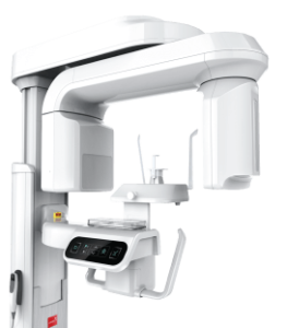
What is a periapical dental x-ray?

It’s a digital x-ray image of 1–3 teeth with high-resolution visualization of roots, canals, and surrounding bone.

How many times can you get a periapical x-ray?

The procedure can be repeated as often as needed for treatment monitoring, since the radiation dose is minimal.

Can you get a periapical x-ray during pregnancy?

In the first trimester – only when absolutely necessary. In the second and third trimesters, it’s permitted with additional protection (lead apron).

What's the difference between periapical and panoramic x-rays?

Periapical x-rays show 1–3 teeth in detail, while panoramic x-rays (also called OPG or orthopantomogram) capture the entire jaw but with less detail.

Can I get a copy of my x-ray?

Yes, the digital image can be saved to a flash drive, disc, or sent via email for future use.

Reviews of targeted dental imaging at Med-Deo

Leave a review
5

Based on reviews

Based on reviews

Андрій

2026-06-11

Робили прицільний знімок перед лікуванням складного зуба. Усе виконали швидко, а лікар одразу пояснив результати.

Татьяна Минко

2026-05-06

Все чудово!

tages tag

2026-04-19

Супер. Швидко, професійно. І головне у вихідні

Ростислав

2026-03-24

Робили прицільний знімок перед лікуванням. Все швидко і без дискомфорту. Лікар одразу показав проблему. Дуже зручно.

Роман

2026-02-20

Зручно, що все на місці, не потрібно їхати окремо в інший центр. Процедура займає кілька хвилин.

Павло

2026-01-14

Робили прицільний знімок перед лікуванням. Швидко, без черг. Одразу пояснили, що видно на знімку.

Show more Hide

What is a Periapical Dental X-Ray?

A periapical dental x-ray is a digital radiographic image that provides a clear and detailed picture of 1–3 teeth along with their roots, root canals, periodontal ligament, and surrounding bone tissue. In dental practice, this method is also known as digital radiography, intraoral x-ray, or simply a dental x-ray.

The examination is performed using a digital sensor that replaces traditional x-ray film. The image appears on the computer screen almost instantly, allowing the dentist to immediately assess the tooth’s condition and make a decision about treatment.

The main difference between a periapical x-ray and a panoramic one lies in the level of detail. Periapical radiography focuses on a small area—specific teeth and their immediate surroundings—whereas a panoramic x-ray (OPG) covers the entire jaw but with less precision. That’s exactly why periapical x-rays are indispensable for diagnosing root canals, hidden cavities, and inflammatory processes.

Why You Need a Periapical X-Ray: Indications for the Procedure?

A periapical x-ray is ordered when a routine examination isn’t sufficient. It helps the dentist see hidden problems and make an informed treatment decision.

First, periapical x-rays are used for diagnostics. They help identify interproximal cavities (between teeth), inflammation under fillings or crowns, cysts, granulomas, periapical periodontitis, pulpitis, as well as cracks or root fractures that are impossible to see with the naked eye.

Second, the x-ray is needed for treatment monitoring. It allows assessment of root canal obturation quality, verification of working length during endodontic treatment, and tracking the healing process after tooth extraction or surgical procedures.

Third, periapical x-rays are used for treatment planning—before implant placement, prosthetic work, wisdom tooth extraction, or beginning orthodontic treatment.

Main Indications for Periapical X-Rays:

  • Unexplained tooth pain
  • Deep or interproximal cavities
  • Suspected periapical infection
  • Dental trauma
  • Before root canal treatment
  • Post-treatment evaluation after endodontics
  • Impacted wisdom teeth
  • Bone loss assessment in periodontal disease

A periapical x-ray is a simple and accurate way to get a complete picture of your dental health and avoid treatment errors.

Need Diagnostics?

Appointment by phone:

(044) 394-90-94

Sign up
icon icon
дагностика 1

What a Periapical X-Ray Shows: Problems That Can Be Identified

A periapical x-ray helps the dentist see what’s invisible during a routine clinical examination and accurately assess the condition of teeth and surrounding bone tissue.

It can reveal:

  1. Dental caries—especially interproximal decay or recurrent caries under fillings that are hidden from view; the x-ray shows radiolucent (dark) areas of enamel and dentin breakdown.
  2. Pulpitis and apical periodontitis—inflammation of the pulp or periapical tissues; visible as widening of the periodontal ligament space and periapical radiolucency at the root apex.
  3. Periapical cysts and granulomas—rounded radiolucent lesions of various sizes at the root apex.
  4. Root canal filling quality—whether canals are adequately obturated, if there are voids or overfilled material extending beyond the apex.
  5. Bone resorption—characteristic of periodontal disease or chronic inflammation.
  6. Root fractures and cracks—fracture lines visible after trauma.
  7. Implant and post condition—checking osseointegration and absence of periapical pathology.
  8. Impacted teeth—teeth that haven’t properly erupted, such as horizontally positioned wisdom teeth.

Such x-rays allow for timely diagnosis, treatment planning, and prevention of complications.

How to Get a Periapical X-Ray

How do you get a periapical x-ray? The procedure is simple and painless, takes just a few minutes, and provides a detailed image of the tooth and surrounding tissues.

  1. Preparation—The patient sits in the dental chair and removes metal jewelry (earrings, necklaces) that could create artifacts on the x-ray.
  2. Protection—A lead apron is placed over the patient’s body, shielding the torso and thyroid gland.
  3. Sensor placement—The dentist positions a small digital sensor intraorally next to the tooth; the patient bites on a positioning device.
  4. Exposure—The x-ray tube head is positioned extraorally (near the cheek), and a brief exposure of 1–2 seconds is taken; it’s important to remain still.
  5. Image—The image instantly appears on the computer monitor and can be magnified for detailed analysis.

Procedure time: 2–3 minutes per x-ray.

Sensation: Completely painless; the only discomfort is having the sensor in your mouth.

Number of images: If necessary, multiple x-rays can be taken during one visit—for different teeth or angulations.

Need Treatment?

Schedule an appointment!

Sign up
icon icon
photo

Periapical X-Ray: Safe Diagnostics

The radiation dose from one periapical x-ray is 0.002–0.005 mSv, which is 1000 times less than the annual background exposure and equivalent to 1–2 hours of air travel. Digital radiography reduces the dose by another factor of 10 compared to traditional film.

  • Pregnant patients: First trimester—only when absolutely necessary; second and third trimesters—with double lead shielding.
  • Children: Safe from ages 3–4 years.

Getting a periapical x-ray allows you to quickly and safely assess the condition of teeth, root canals, and surrounding tissues.

Other Dental Imaging Services
icon icon 9 rentgen
We Partner with All Insurance Companies

On this page Contents of the page

icon

Dental services