Special offer! Comprehensive professional teeth cleaning for only 1699 UAH.
Special offer! Comprehensive professional teeth cleaning for only 1699 UAH.
Special offer! Comprehensive professional teeth cleaning for only 1699 UAH.
Special offer! Comprehensive professional teeth cleaning for only 1699 UAH.
Special offer! Comprehensive professional teeth cleaning for only 1699 UAH.
Special offer! Comprehensive professional teeth cleaning for only 1699 UAH.
Special offer! Comprehensive professional teeth cleaning for only 1699 UAH.
Special offer! Comprehensive professional teeth cleaning for only 1699 UAH.
Special offer! Comprehensive professional teeth cleaning for only 1699 UAH.
Special offer! Comprehensive professional teeth cleaning for only 1699 UAH.
Sign up
 (044) 394 90 94
Sign up
(044) 394 90 94
Doctors


 Prices
Blog
Contacts
Prices
Blog
Contacts
пр.Лобановського, 130, Київ, Україна
Temporarily closed
Opening soon
78A, Irpinska St., Kyiv, Ukraine
Temporarily closed
Opening soon
32A, Heroiv Dnipra St., Kyiv, Ukraine
Temporarily closed
Opening soon
9B, Yevhena Chykalenko St. (Pushkinska), Kyiv, Ukraine
Temporarily closed
Opening soon
(044) 394 90 94
Back
Dentistry
General Dentistry
Dental surgery
Dental Implants
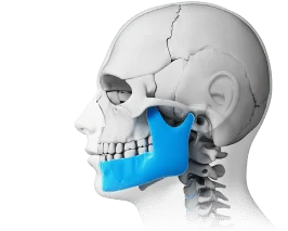
Maxillofacial Surgery
Dental prosthetics
Cosmetic dentistry
Professional Dental Hygiene

Periodontics
Orthodontic dentistry
Gnathology & TMJ Therapy
Pediatric Dentistry
Dental X-Rays
Search on the site


Popular requests
Teeth whitening
Treatment of caries
Wisdom tooth extraction
Bracket system
Dental veneers
Tooth restoration
Pediatric dentist
Consultation with a dentist-therapist
Search results by: «».
Unfortunately, no results were found for your search: “Pediatric Endocrinologist”, nothing was found.
You may find these links useful:
Clinics "MED-DEO"
пр.Лобановського, 130, Київ, Україна
Temporarily closed
Opening soon
78A, Irpinska St., Kyiv, Ukraine
Temporarily closed
Opening soon
32A, Heroiv Dnipra St., Kyiv, Ukraine
Temporarily closed
Opening soon
9B, Yevhena Chykalenko St. (Pushkinska), Kyiv, Ukraine
Temporarily closed
Opening soon
Services
DentistryGeneral Dentistry
Dental surgery
Dental Implants
Maxillofacial Surgery
Dental prosthetics
Cosmetic dentistry
Professional Dental Hygiene
Periodontics
Orthodontic dentistry
Gnathology & TMJ Therapy
Pediatric Dentistry
Dental X-Rays

")
An impacted tooth and ectopia are the two most common problems associated with wisdom teeth. In short: the tooth either cannot erupt at all or grows in the wrong direction.
According to statistics, more than half of people experience difficult eruption of “third molars.” And this is not accidental. Wisdom teeth erupt last—between ages 17–25, when the jaw is already formed and there is often simply not enough space. Because of this, they become stuck in the bone, partially emerge, or grow at an angle, pressing on adjacent teeth.
At first glance, the problem may not cause concern. But over time, impaction and ectopia can cause pain, gum inflammation, cavities in neighboring teeth, and even bite misalignment. This is precisely why dentists recommend not ignoring such situations.

What Is an Impacted Tooth
What is an impacted tooth? It is a condition when a tooth has fully formed but cannot erupt normally and remains under the gum or inside the jawbone.
Simply put: the tooth exists, but it is “stuck” and does not come out.
Tooth impaction can manifest differently:
- produce no symptoms and be discovered accidentally on an X-ray
- cause periodic pain or pressure in the jaw
- cause swelling and redness of the gums
- provoke discomfort during chewing
- sometimes be accompanied by unpleasant odor due to inflammation around the tooth
Why Wisdom Teeth Most Often Become Impacted
An impacted wisdom tooth is the most common case of this pathology. The reason is simple:
- they erupt last
- often do not have enough space
- grow at an incorrect angle or press against the “sevens” (second molars)
As a result, the “eights” (third molars) either remain under the gums or emerge partially, creating ideal conditions for inflammation.
What Is Tooth Ectopia
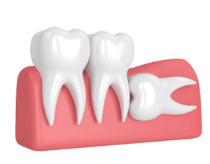
Tooth ectopia is a violation of the tooth’s position in the dental arch. That is, the tooth has erupted but is not in its proper place or grows at an incorrect angle.
In practice, this may look like:
- tooth shifted toward the cheek or tongue
- tilted forward or backward
- rotated around its axis
- protruding from the row or “hiding” behind neighboring teeth
- growing toward the palate
Any tooth can be ectopic—from an incisor to a molar. But most often dentists encounter ectopia of:
- wisdom teeth
- canines
- front teeth
The problem is not only aesthetic. Incorrect tooth position complicates hygiene, promotes cavities, injures the mucosa, and can affect the bite.
Difference Between Tooth Impaction and Ectopia
Although these terms are often confused, they mean different problems.
Tooth impaction is an eruption disorder. The tooth has formed but has not emerged and remains under the gum or in the bone.
Tooth ectopia is a position disorder. The tooth has erupted but grew incorrectly and occupies the wrong position in the dental arch.
It is important to understand that these conditions often combine. For example:
- an impacted wisdom tooth may lie horizontally in the bone
- or be tilted and press on an adjacent tooth
- partially erupt and simultaneously have an incorrect growth direction
In such cases, we speak of an impacted ectopic tooth (when ectopia and impaction occur simultaneously), and precisely such situations most often require surgical treatment.
Why Tooth Impaction and Ectopia Occur: Main Causes
The appearance of these problems is not accidental. They have clear anatomical and genetic causes.
Evolutionary Changes in the Jaw
During evolution, the size of the human jaw decreased, while the number of teeth remained the same—32. As a result, there is often simply not enough space for wisdom teeth, so they:
- become stuck in the bone
- grow at an angle
- erupt partially or do not emerge at all
Genetic Factors
Very often impaction and ectopia have a hereditary basis. A person may inherit:
- a small jaw
- large-sized teeth
- peculiarities of tooth bud growth
This creates an anatomical “space conflict” even before the appearance of permanent teeth.
Intrauterine Development Disorders
Factors during pregnancy also affect tooth formation:
- deficiency of vitamins and minerals
- infectious diseases in the mother
- disruption of tooth bud development
All this can change the direction of tooth growth even before birth.
Acquired Causes
In addition to heredity, external factors also play a significant role:
- early removal of baby teeth without space maintenance
- jaw injuries
- harmful habits (thumb sucking, prolonged pacifier use)
- improper breastfeeding and disruption of bite formation
All this gradually changes tooth position and creates conditions for ectopia and impaction.
Symptoms and Signs of Impacted and Ectopic Teeth
Impacted and ectopic teeth can manifest differently—from complete absence of symptoms to pronounced pain and inflammation. This is precisely why many patients ignore the problem for a long time until it transitions to an acute phase.
Signs of an Impacted Tooth
Impacted teeth may not cause concern for a long time, but most often the following symptoms appear:
- gum pain that intensifies during chewing or opening the mouth
- swelling and redness of the gum in the problem tooth area
- sensation of pressure or “bursting” in the jaw
- appearance of a so-called “hood”—an overhanging section of gum over a partially erupted wisdom tooth
- unpleasant breath odor due to bacterial accumulation under the gums
Such symptoms especially often occur with wisdom tooth impaction.
Signs of Tooth Ectopia
An ectopic tooth is usually noticeable immediately, as it changes the appearance of the smile and position of the dental arch. Main manifestations:
- cosmetic defect—the tooth “sticks out,” protrudes, or stands crookedly
- injury to the oral mucosa—biting of the cheek, tongue, gums
- discomfort or pain during chewing
- bite misalignment
- displacement of neighboring teeth
- speech problems with pronounced anomalies
- difficulty with hygiene—accumulation of plaque and tartar in hard-to-reach places
Even if pain is absent, ectopia gradually creates conditions for serious dental problems.
What Problems and Complications May Arise
Ignoring impacted and ectopic teeth is one of the most common causes of chronic inflammations in the oral cavity. Without treatment, the situation almost always worsens.
Inflammatory Processes
The most common complications:
- pericoronitis—inflammation of tissues around the wisdom tooth
- stomatitis
- periodontitis and gum inflammation
Such conditions are accompanied by pain, swelling, unpleasant odor, and can transition to chronic form.
Infectious Complications
With prolonged inflammation, serious consequences are possible:
- formation of a follicular cyst around the impacted tooth
- gum abscess
- phlegmon—diffuse purulent inflammation of soft tissues that poses a real threat to life and requires emergency care
Damage to Neighboring Teeth
An ectopic or impacted wisdom tooth can press on the “seven” (second molar), leading to:
- development of cavities in the contact zone
- enamel destruction
- resorption (dissolution) of neighboring tooth roots
Orthodontic Problems
Constant pressure and incorrect growth direction cause:
- displacement of the entire dental arch
- tooth crowding
- formation of incorrect bite
- deterioration of orthodontic treatment results
Neurological Complications
In complex cases, the following are possible:
- irritation or inflammation of the trigeminal nerve
- pain in the temporomandibular joint
- radiating pain to the ear, temple, neck
This is precisely why dentists recommend not waiting for complications but solving the problem at an early stage.
Diagnosis of Tooth Impaction and Ectopia
Accurate diagnosis is a key stage of proper treatment. Visual examination is often insufficient, especially if the tooth is located inside the bone.
Dentist Examination
During examination, the doctor can:
- notice ectopia with the naked eye
- assess the condition of gums, bite, tooth position
- suspect impaction by indirect signs
But visualization is always required to confirm the diagnosis.
Radiography
A periapical X-ray allows:
- seeing the position of the impacted tooth
- assessing the growth direction
- identifying inflammatory changes around the root
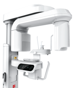
Orthopantomogram (Panoramic Image)
A panoramic image of the jaws provides an overall picture:
- location of all wisdom teeth
- root condition
- interaction of teeth with each other
- presence of hidden pathologies
CBCT—Cone Beam Computed Tomography
This is the most informative diagnostic method. It creates a three-dimensional image and allows:
- precisely determining the position of the impacted tooth
- assessing proximity to the mandibular canal and maxillary sinus
- seeing contact with roots of neighboring teeth
- properly planning surgical intervention
Treatment Methods for Impacted and Ectopic Teeth
Treatment tactics are selected individually. Everything depends on the tooth’s position, presence of symptoms, and risks of complications. Dentistry uses three main strategies.
1. Observation
Used in cases when:
- the impacted tooth does not cause pain
- there is no inflammation
- it does not press on neighboring teeth
- orthodontic problems are absent
The patient undergoes regular examinations and X-ray monitoring. But even with observation, it is important to understand: the situation can change over time.
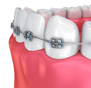
2. Orthodontic Treatment
Applied when the tooth has potential for correct position. Most often this concerns canines or front teeth.
The essence of the method is pulling the tooth into the dental arch using braces or special orthodontic appliances. The process requires time but allows preserving one’s own tooth.
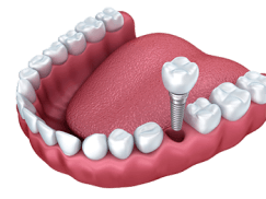
3. Surgical Removal
The most common option for impacted and ectopic wisdom teeth. This method is applied when the tooth creates health risks or already causes complications.
How Atypical Removal of an Impacted or Ectopic Tooth Proceeds
Many patients are frightened by the word “surgery,” but in practice the procedure is clearly regulated and proceeds under complete anesthesia.
Stage 1. Anesthesia
Local anesthesia or sedation is performed. The patient does not feel pain, only pressure and touch.
Stage 2. Creating Access
The surgeon:
- makes an incision in the mucous membrane
- reflects the gingival flap
- uses a drill to remove a small section of bone to expose the tooth crown
Stage 3. Tooth Removal
Tactics depend on root structure:
- with simple anatomy, the tooth is removed entirely
- with complex root system, segmentation is performed—the tooth is sawed into parts and removed in stages
This reduces tissue trauma.
Stage 4. Socket Processing
The doctor:
- smooths sharp bone edges
- rinses the wound with antiseptic solutions
- checks complete removal of fragments
Stage 5. Suturing
The socket is closed with a mucous flap and sutures are placed. This accelerates healing and reduces infection risk.
Contraindications to Removal of Impacted Teeth
Before surgery, the doctor necessarily assesses the patient’s overall health condition.
Absolute Contraindications
Surgery is postponed or canceled with:
- severe cardiovascular diseases in decompensation stage
- blood clotting disorders (for example, hemophilia)
- acute infectious diseases, including COVID-19
Relative Contraindications
Removal is temporarily postponed with:
- exacerbation of chronic diseases
- first and third trimesters of pregnancy
- uncontrolled arterial hypertension
- diabetes mellitus in decompensation stage
After condition stabilization, surgery can be performed safely.
Rehabilitation After Removal: What to Expect
The postoperative period proceeds differently for each patient. It depends not only on surgery complexity but also on individual body characteristics.
Normal Symptoms in the First 3–7 Days
May be observed:
- pain (controlled by pain medications)
- cheek swelling reaching peak on days 2–3
- difficulty opening the mouth
- slight temperature increase to 37.5°C
- general weakness
This is a normal body reaction to surgical intervention.
Recommendations After Removal
For rapid healing, it is important to:
- apply cold compresses in the first 24 hours
- consume soft, warm food
- avoid physical activity
- not rinse the mouth aggressively for the first 2 days
- take medications prescribed by the doctor
Follow-up Examinations
Usually:
- the next day telephone contact with the surgeon’s assistant is conducted
- if necessary—examination at the clinic
- suture removal after 7–10 days
Alarming Symptoms
Immediately consult a doctor if the following appear:
- severe swelling that does not decrease for more than 3 days
- temperature above 38°C
- prolonged bleeding
- numbness that does not pass
Possible Complications After Surgery
Any surgical intervention has certain risks, especially with complex tooth anatomy.
Possible complications:
- damage to the mandibular nerve—temporary numbness of lip, chin, or tongue
- damage to the maxillary sinus when removing upper wisdom teeth
- jaw fracture (extremely rare complication)
- injury to neighboring teeth
- alveolitis—socket inflammation
- pushing the root into the maxillary sinus or sublingual area
Important: with an experienced oral and maxillofacial surgeon, these risks are minimal thanks to accurate diagnostics and proper surgical technique.
Questions and Answers (FAQ)
Is it always necessary to remove an impacted tooth?
No. If the tooth does not cause complications and does not create risks—observation is possible.
Is it painful to remove an impacted wisdom tooth?
No. The procedure is performed under anesthesia, pain is absent.
How long does removal of an ectopic tooth take?
On average from 20 to 60 minutes depending on case complexity.
Can ectopia be corrected without removal?
Yes, in some cases orthodontic treatment is possible.
What will happen if an impacted wisdom tooth is not removed?
The risk of inflammation, cysts, damage to neighboring teeth, and bite misalignment increases.
Can an impacted tooth erupt on its own?
Sometimes—yes, but after ages 20–25 the probability significantly decreases.
Written by Anton Semenov, DDS Oral Surgeon & Implantologist


")

Make an appointment
You can make an appointment by filling out the application form on the website, as well as using instant messengers:
Make an appointment
You can make an appointment by filling out the application form on the website, as well as using instant messengers:


Make an appointment easily using chatbots:
Also, call the numbers:
Modern dentistry and medical centre "MED-DEO"
Choose a clinic whose location is more convenient for you.
Kyiv, Holosiivska/Demiivska metro station
130 Lobanovskoho Avenue
")
")
")
")
Working hours:
Mon - Sun: 8:00 AM - 9:00 PM
No days off!
Call the numbers:
How to get to us? Create a route
Kyiv, Akademistechko metro station
78A, Irpinska str.


Working hours:
Mon-Sun: 8.00-21.00
Without days off!
Call the numbers:
How to get to us?
Create a route
Kyiv, Heroiv Dnipra metro station
32A, Heroiv Dnipra str.


Working hours:
Mon-Sun: 8.00-21.00
Without days off!
Call the numbers:
How to get to us?
Create a route
Kyiv, Teatralna metro station
9B, E. Chykalenko (Pushkinska) str.


Working hours:
Mon-Sat: 8.00 - 21.00. Sun: by appointment.
Without days off!
Call the numbers:
How to get to us?
Create a route
Відгук від клієнта
0:00
0:00

Your email will not be published on the site.