
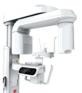
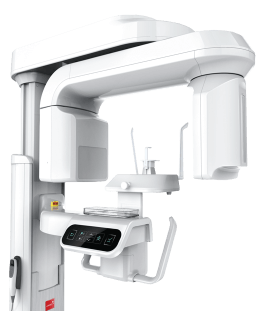
Orthopedic dentistry and prosthetics performed under a microscope begin with a consultation. The dentist examines the teeth and gums, and assesses the bite, the condition of existing fillings, crowns, and abutment teeth, as well as the overall clinical situation. If necessary, X-rays, CT scans, or other diagnostic procedures are prescribed.
The next stage is the preparation of the oral cavity. Before a crown, bridge, or denture is placed, it is essential to treat any tooth decay or gum inflammation, replace inadequate old fillings, and—if required—perform endodontic treatment. An orthopedic restoration must be anchored on a healthy and stable foundation.
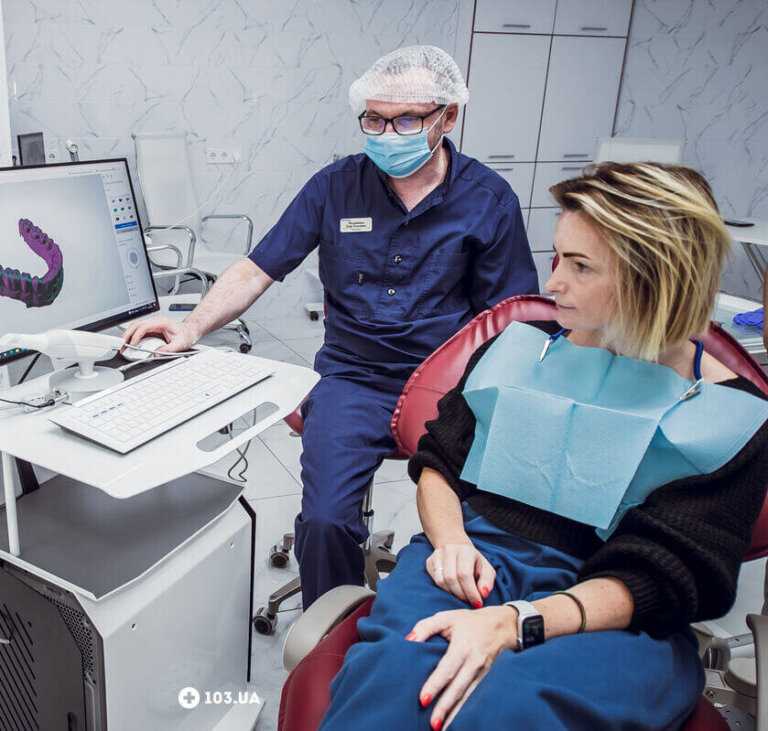
Following preparation, the dentist proceeds to prepare the tooth. In appropriate cases, this stage may be performed under a microscope. Next, impressions are taken or a digital scan is performed to ensure the final restoration is fabricated with maximum precision.
While the permanent crown or other restoration is being crafted in the laboratory, a temporary crown may be placed. This protects the tooth, maintains aesthetics, and ensures a more comfortable waiting period.
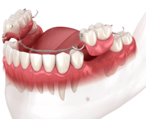
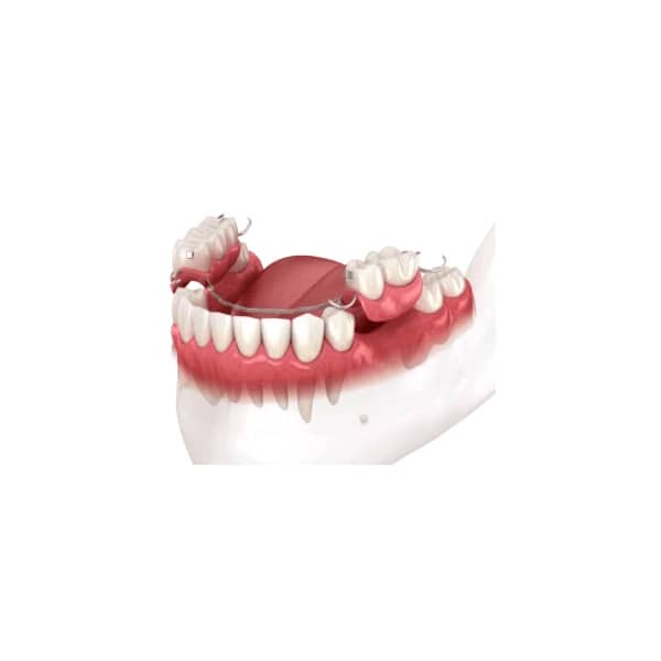
Once the permanent restoration is ready, the dentist performs a try-in to check the fit, color, shape, contact points with adjacent teeth, occlusion (bite), and comfort during chewing. If everything meets clinical standards, the crown, bridge, or prosthesis is permanently fixed in place.
The final stage involves care instructions and follow-up appointments. High-quality prosthetic treatment does not end at the moment of fixation; it is essential to properly maintain the restoration, attend preventive check-ups, and address any discomfort should it arise.
 (044) 394 90 94
 Holosiivska/Demiivska metro station

























Your email will not be published on the site.