Special offer! Comprehensive professional teeth cleaning for only 1699 UAH.
Special offer! Comprehensive professional teeth cleaning for only 1699 UAH.
Special offer! Comprehensive professional teeth cleaning for only 1699 UAH.
Special offer! Comprehensive professional teeth cleaning for only 1699 UAH.
Special offer! Comprehensive professional teeth cleaning for only 1699 UAH.
Special offer! Comprehensive professional teeth cleaning for only 1699 UAH.
Special offer! Comprehensive professional teeth cleaning for only 1699 UAH.
Special offer! Comprehensive professional teeth cleaning for only 1699 UAH.
Special offer! Comprehensive professional teeth cleaning for only 1699 UAH.
Special offer! Comprehensive professional teeth cleaning for only 1699 UAH.
 (044) 394 90 94
  Holosiivska/Demiivska metro station
(044) 394 90 94
Holosiivska/Demiivska metro station
(044) 394 90 94 пр.Лобановського, 130, Київ, Україна
Temporarily closed
Opening soon

78A, Irpinska St., Kyiv, Ukraine
Temporarily closed
Opening soon

32A, Heroiv Dnipra St., Kyiv, Ukraine
Temporarily closed
Opening soon

9B, Yevhena Chykalenko St. (Pushkinska), Kyiv, Ukraine
Temporarily closed
Opening soon

General Dentistry
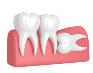
Dental surgery
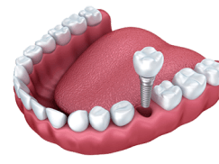
Dental Implants
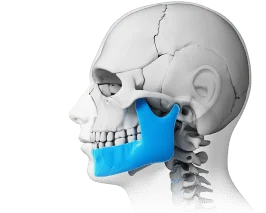
Maxillofacial Surgery
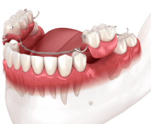
Dental prosthetics
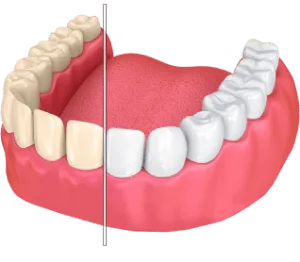
Cosmetic dentistry
Professional Dental Hygiene

Periodontics
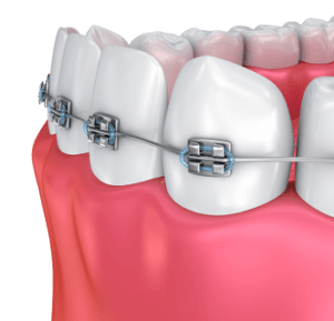
Orthodontic dentistry
Gnathology & TMJ Therapy

Pediatric Dentistry
Dental X-Rays
Orthodontic dentistry
Gnathology & TMJ Therapy
Dental prosthetics
Maxillofacial Surgery
Plastic surgery
Dental surgery
Dental Implants


Popular requests
Teeth whitening
Treatment of caries
Wisdom tooth extraction
Bracket system
Dental veneers
Tooth restoration
Pediatric dentist
Consultation with a dentist-therapist
Search results by: «».
Unfortunately, no results were found for your search: “Pediatric Endocrinologist”, nothing was found.
You may find these links useful:
Clinics "MED-DEO"
пр.Лобановського, 130, Київ, Україна
Temporarily closed
Opening soon
78A, Irpinska St., Kyiv, Ukraine
Temporarily closed
Opening soon
32A, Heroiv Dnipra St., Kyiv, Ukraine
Temporarily closed
Opening soon
9B, Yevhena Chykalenko St. (Pushkinska), Kyiv, Ukraine
Temporarily closed
Opening soon
Services
Dentistry Orthodontic Center Prosthodontic Center MED-DEO Center for Maxillofacial Surgery & Facial Plastics Surgical Dental CenterGeneral Dentistry
Dental surgery
Dental Implants
Maxillofacial Surgery
Dental prosthetics
Cosmetic dentistry
Professional Dental Hygiene
Periodontics
Orthodontic dentistry
Gnathology & TMJ Therapy
Pediatric Dentistry
Dental X-Rays
Orthodontic dentistry
Gnathology & TMJ Therapy
Dental prosthetics
Maxillofacial Surgery
Dental surgery
Dental Implants

Service statistics

More and more clients choose «MED-DEO»

Initial doctor's consultation for free* * subject to treatment in the clinic
Appointment by phone:
(044) 394 90 94One of the primary methods for diagnosing oral health issues is a dental X-ray with an interpretation.
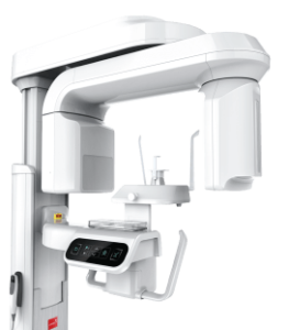
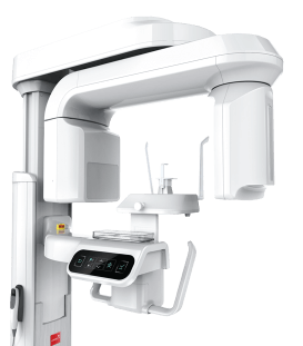
To ensure an accurate diagnosis and the selection of the most effective solution, a tomograph or radiovisiograph is also used. This innovative equipment enables the generation of even a three-dimensional visualization of a specific area.
Price for the interpretation of dental X-rays
Interpretation of dental X-rays at the Med-Deo clinic
The Med-Deo clinic provides X-rays with diagnostic reports and computed tomography results. Innovative, certified, and high-precision equipment is used for dental imaging. You can have your scan performed here and receive an interpretation from experienced radiologists.
The clinic offers a full range of dental services. Should any issues with teeth or gums be detected, the top doctors—professional and experienced dentists at Med-Deo—are ready to provide treatment at reasonable prices.









Reviews of dental X-ray interpretation at Med-Deo
When is a description of dental X-rays necessary?
Not all dental clinics today are equipped to take X-rays, as they may lack the necessary X-ray or digital imaging equipment.
If your chosen clinic does not have this equipment, it is best to arrive for your appointment with the imaging results already in hand. X-rays allow for a highly accurate assessment of the condition of tissues, teeth, and roots without causing the patient pain or discomfort. Furthermore, this diagnostic method enables the timely detection of serious conditions, allowing treatment to begin immediately. An orthopantomogram—essentially a “profile” of the dentofacial system—or a dental CT scan with a radiologist’s report is particularly useful. Since the general condition of the oral cavity does not change rapidly, a single examination is usually sufficient for treatment planning. The “Med-Deo” clinic in Kyiv offers the following diagnostic services, including a professional report:
- Panoramic X-ray;
- CT scan of a single jaw;
- CT scan of both jaws;
- CT scan of a specific segment.

The results can be provided as an image on specialized film or on digital media (in the case of a CT scan).


What can be learned from the description of a dental X-ray?
This health-safe scan provides the doctor with information regarding the presence or absence of issues and pathologies.
At our dental clinic, the interpretation of dental CT scans and X-rays is performed by experienced radiologists. These diagnostic methods enable the timely detection of:
- Cracks and root fractures, as well as the assessment of the aftereffects of trauma. An X-ray is mandatory if the patient exhibits noticeable gum inflammation, severe pain during chewing or biting, or tooth mobility.
- Abnormalities of the temporomandibular joint, jawbone, and bite (occlusion).
- Diseases. Periodontitis: a condition requiring immediate medical intervention due to bone tissue atrophy, gum bleeding, inflammation and tenderness, and tooth mobility; Periodontitis (apical): the development of cysts or granulomas at the root apex, leading to the formation of fistulas, abscesses, and ultimately, tooth loss.
- Caries in hidden areas of the oral cavity. X-rays allow for the detection of carious lesions beneath crowns and behind fillings.
- Abscesses, tumors, and other growths.
- Cracks and root fractures, as well as the assessment of the aftereffects of trauma. An X-ray is mandatory if the patient exhibits noticeable gum inflammation, severe pain during chewing or biting, or tooth mobility.
- Abnormalities of the temporomandibular joint, jawbone, and bite (occlusion).
- Diseases. Periodontitis: a condition requiring immediate medical intervention due to bone tissue atrophy, gum bleeding, inflammation and tenderness, and tooth mobility; Periodontitis (apical): the development of cysts or granulomas at the root apex, leading to the formation of fistulas, abscesses, and ultimately, tooth loss.
- Caries in hidden areas of the oral cavity. X-rays allow for the detection of carious lesions beneath crowns and behind fillings.
- Abscesses, tumors, and other growths.
Visiography, X-rays, and 2D and 3D models of the teeth and temporomandibular joint make it possible to significantly improve the quality of dental care. These images also allow for monitoring treatment effectiveness and assessing the feasibility of implant placement and orthopedic procedures.
Do you have any remaining questions?
Book an appointment with the doctor!


Dental services
General Dentistry
Dental surgery
Dental Implants
Maxillofacial Surgery
Dental prosthetics
Cosmetic dentistry
Professional Dental Hygiene
Periodontics
Orthodontic dentistry
Gnathology & TMJ Therapy
Pediatric Dentistry
Dental X-Rays
Dental Tourism
Відгук від клієнта


Ірина
2026-06-06
Робила рентгенівські знімки перед лікуванням. Лікар детально пояснив кожен проблемний зуб і відповів на всі мої запитання.
Олена
2026-05-07
Звернулась для розшифровки знімків зубів. Лікар дуже доступно пояснив кожну проблему і показав усе на екрані. Після консультації стало зрозуміло, що потрібно лікувати.
Ірина
2026-02-12
Робили опис знімка перед лікуванням. Лікар детально пояснив кожен момент, стало зрозуміло, що відбувається.
Валерія
2025-10-10
Була на консультації у лікаря з КТ, він описа всі проблеми по всім зубам, та склав лікування. Дуже сподобалося відношення і лікаря і адміністраторів. Рекомендую!
Олена
2025-09-02
Робила рентген зубів у клініці – все швидко, безболісно та якісно. Знімок відразу віддали на руки й пояснили результат.
Іван
2025-07-05
Робив КТ щелепи для планування імплантації. Отримав дуже детальний опис, лікар пояснив кожну зону, звернув увагу навіть на ті моменти, яких я не очікував. Після консультації залишився з чітким розумінням, що і коли потрібно робити. Дякую!
Олена
2024-10-22
Робила ОПТГ (панорамний знімок). Дуже сподобалось відношення персоналу. Усе ввічливо, швидко. Гарна якість. Сервіс на висоті. Рекомендую.
Володимир
2022-02-10
Швидко зробили знімок, підсказали на які зуби потрібно звернути увагу. Персонал ввічливий та пунктуальний! Рекомендую!